Apex Scientific

21+ access.
Research use only.

Before entering, confirm that you meet the age requirement and understand the research-use limitations that apply to every Apex product.

Leave site

Your acknowledgement is remembered on this device for 30 days.

Research use only Clear product information Free shipping on orders over $200 20% launch offer · Use code APEX101

Compound guide

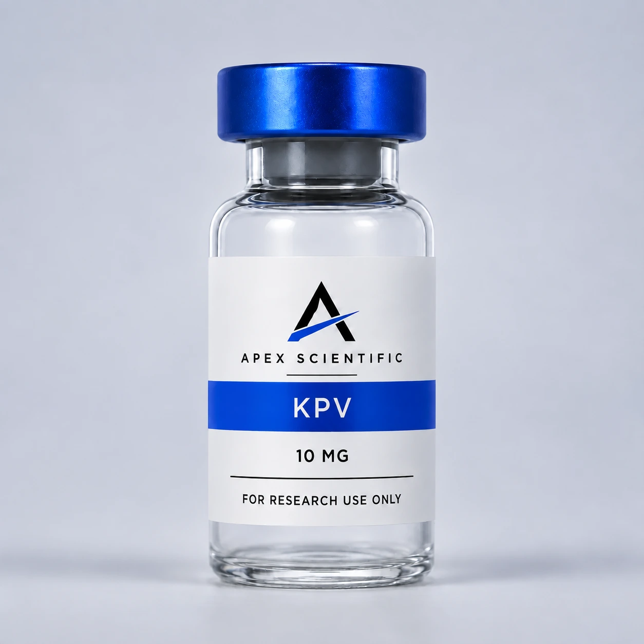
KPV

A pathway-focused guide to molecular mechanisms, published evidence, and the limits of interpretation.

Overview

A Summary:KPV

KPV is the three-amino-acid sequence lysine-proline-valine, corresponding to the C-terminal 11–13 fragment of alpha-melanocyte-stimulating hormone.

Cell studies use KPV to examine melanocortin-related signaling, intracellular calcium responses, and inflammatory-response pathways without assuming that it reproduces the full parent peptide.

The published evidence is primarily cellular and preclinical. A molecular response in cultured cells does not establish a safe or effective human outcome, and controlled human efficacy has not been established.

Research model

Cellular & MolecularHow KPV Is Studied

These receptors, enzymes, and signaling networks define the primary laboratory questions associated with this research material.

01Parent-peptide fragment

Melanocortin-related signaling

At the cellular or molecular level

KPV corresponds to the final three residues of alpha-MSH. Cell experiments compare its responses with alpha-MSH and related ACTH fragments to determine which signals depend on the full peptide.

02Keratinocyte model

Intracellular calcium response

At the cellular or molecular level

A cultured-cell study reported rapid intracellular-calcium responses to KPV under specific experimental conditions while not detecting a cyclic-AMP increase in the keratinocyte models tested.

03Preclinical hypothesis

Inflammatory-response network

At the cellular or molecular level

KPV is used in laboratory models to examine signaling associated with cellular stress and inflammatory responses. The downstream route may vary by model and remains under investigation.

Research interpretation

How to read the pathway.

01

Parent-peptide fragment

KPV corresponds to the final three residues of alpha-MSH.

02

Keratinocyte model

A cultured-cell study reported rapid intracellular-calcium responses to KPV under specific experimental conditions while not detecting a cyclic-AMP increase in the keratinocyte models tested.

03

Preclinical hypothesis

KPV is used in laboratory models to examine signaling associated with cellular stress and inflammatory responses.

Evidence boundary

The published evidence is primarily cellular and preclinical. A molecular response in cultured cells does not establish a safe or effective human outcome, and controlled human efficacy has not been established.
Read the evidence

Sources BehindThis Guide

KPV signaling in human keratinocyte cells (opens in a new tab)L-lysyl-L-prolyl-L-valine identity record (opens in a new tab)

One important note

Educational Notice

KPV evidence is primarily cellular and preclinical. Published cell responses do not establish safety, efficacy, or a predictable human outcome.

This guide summarizes molecular mechanisms, published evidence, and evidence limits in everyday language. It is educational information, not medical advice or instructions for personal use. Products offered on this site are intended exclusively for laboratory research and are not for human or veterinary use.